


From the article you will learn the features of varicose veins of the pelvis in women - this is a deformation of the veins of the pelvic area with impaired blood flow to the internal and external genitalia.
Main information
In the literature, varicose veins of the pelvis are also called "pelvic congestion syndrome", "varicocele in women", "chronic pelvic pain syndrome". The prevalence of varicose veins in the pelvis increases in proportion to age: from 19. 4% in girls under 17 to 80% in perimenopausal women. Most often the pathology of the pelvic veins is diagnosed in the reproductive period in patients in the age group 25-45 years.
In the majority of cases (80%) varicose transformation affects the ovarian veins and is extremely rare (1%) in the veins of the broad ligament of the uterus. According to modern medical approaches, the treatment of VVMT should be performed not so much from the point of view of gynecology, but first of all from the point of view of phlebology.
Causes of pathology
By varicose veins of the pelvic organs in women, doctors understand a change in the structure of the vessel walls, characteristic of other types of disease - weight loss, followed by stretching and the formation of "pockets" inside which the blood stagnates. Extremely rare are cases where only the vessels of the pelvic organs are affected. In about 80% of patients, along with this form, there are signs of varicose veins of the inguinal veins, vessels of the lower extremities.
The incidence of varicose veins of the pelvis is most pronounced in women. This is due to anatomical and physiological features that suggest a tendency to weaken the venous walls:
- hormonal fluctuations, including those related to the menstrual cycle and pregnancy;
- increased pressure in the small pelvis, which is characteristic of pregnancy;
- periods of more active filling of the veins with blood, including cyclic menstrual periods, during pregnancy, as well as during sex.
All these phenomena belong to the category of factors that provoke varicose veins. And they occur exclusively in women. The largest number of patients face varicose veins of the pelvis during pregnancy, as there is a simultaneous layering of provoking factors. According to statistics, in men, varicose veins of the pelvis are 7 times less common than among the fairer sex. They have a more diverse set of provoking factors:
- hypodynamia - long-term maintenance of low physical activity;
- increased physical activity, especially weightlifting;
- obesity;
- lack of enough fiber in the diet;
- inflammatory processes in the organs of the genitourinary system;
- sexual dysfunction or clear refusal of sex.
Genetic predisposition can also lead to pathology of the plexuses located inside the pelvis. According to statistics, varicose veins of the perineum and pelvic organs are most often diagnosed in women whose relatives have suffered from this disease. The first changes in them can be observed in adolescence during puberty.
The greatest risk of developing inguinal varicose veins in women with pelvic vascular involvement is observed in patients with venous pathology in other parts of the body. In this case we are talking about congenital weakness of the veins.
Etiopathogenesis
Proctologists believe that the following main causes always contribute to the appearance of VVP: valve insufficiency, venous obstruction and hormonal changes.
Pelvic venous congestion syndrome can develop due to congenital absence or insufficiency of venous valves, which was revealed by anatomical studies in the last century, and modern data confirm this.
It has also been found that in 50% of patients varicose veins are genetic in nature. FOXC2 is one of the first genes identified to play a key role in the development of VVP. The relationship between disease development and gene mutations (TIE2, NOTCH3), thrombomodulin levels and transforming growth factor β type 2 has now been determined. These factors contribute to changes in the structure of the valve itself or the venous wall - all leading to structural damageon the valve; varicose veins, which causes a change in valve function; to progressive reflux and eventually to varicose veins.
An important role in the development of the disease can be played by connective tissue dysplasia, the morphological basis of which is a decrease in the content of different types of collagen or a violation of the ratio between them, which leads to a decrease in the strength of the veins. .
The frequency of VVP is directly proportional to the amount of hormonal changes that are particularly pronounced during pregnancy. In pregnant women, the capacity of the pelvic veins increases by 60% due to the mechanical compression of the pelvic vessels by the pregnant uterus and the vasodilating effect of progesterone. This venous dilation lasts one month after birth and can cause venous valve insufficiency. In addition, during pregnancy, the mass of the uterus increases, its positional changes occur, which causes dilation of the ovarian veins, followed by venous congestion.
Risk factors also include endometriosis and other inflammatory diseases of the female reproductive system, estrogen therapy, adverse working conditions for pregnant women, which include heavy physical labor and prolonged forced position (sitting or standing) during the working day.
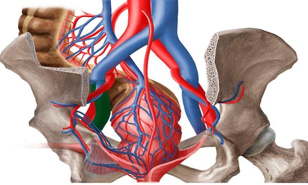
The formation of varicose veins in the pelvis is also facilitated by the anatomical features of the outflow from the pelvic veins. The diameter of the ovarian veins is usually 3-4 mm. The long and thin ovarian vein flows into the left renal vein on the left and into the inferior vena cava on the right. The left renal vein is usually located in front of the aorta and behind the superior mesenteric artery. The physiological angle between the aorta and the superior mesenteric artery is approximately 90 °.
This normal anatomical position prevents compression of the left renal vein. The average angle between the aorta and the superior mesenteric artery in adults is 51 ± 25 °, in children - 45, 8 ± 18, 2 ° in boys and 45, 3 ± 21, 6 ° in girls. In the case of a decrease in the angle from 39, 3 ± 4, 3 ° to 14, 5 °, aorto-mesenteric compression or hazelnut crusher syndrome occurs. This is the so-called anterior, or true, nutcracker syndrome, which has the greatest clinical significance. Posterior hazelnut crusher syndrome occurs in rare cases in patients with retroaortic or annular disposition of the distal left renal vein. Obstruction of the proximal venous bed causes an increase in the pressure in the renal vein, which leads to the formation of renoovarian reflux in the left ovarian vein with the development of chronic pelvic venous insufficiency.
May-Turner syndrome - compression of the left common iliac vein by the right common iliac artery - also serves as one of the etiological factors for varicose veins in the pelvis. It occurs in no more than 3% of cases, it is more common in women. Currently, due to the introduction of radiation and endovascular imaging methods, this pathology is being detected more and more often.
Classification
Varicose veins are divided into the following forms:
- The main type of varicose veins: enlargement of the blood vessels of the pelvis. The reason is valve insufficiency of 2 types: acquired or congenital.
- The secondary form of thickening of the pelvic veins is diagnosed exclusively in the presence of pathologies in terms of gynecology (endometriosis, neoplasms, polycystosis).
Varicose veins of the pelvis develop gradually. In medical practice there are several main stages in the development of the disease. They will vary depending on the presence of complications and the spread of the disease:
- First degree. Changes in the structure of the valves of the ovarian veins can occur due to hereditary causes or be acquired. The disease is characterized by an increase in the diameter of the veins up to 5 mm. The left ovary has a pronounced enlargement in the outer parts.
- Second specialty. This degree is characterized by the spread of pathology and damage to the left ovary. The veins in the uterus and right ovary can also be dilated. The diameter of the extension reaches 10 mm.
- Third degree. The diameter of the veins increases to 1 cm. The dilation of the veins is observed on the right and left ovaries equally. This stage is due to pathological phenomena of gynecological nature.
It is also possible to classify the disease according to the underlying cause of its development. There is a primary stage in which the dilation is caused by a malfunction of the venous valves, and a secondary stage, which is a consequence of chronic female diseases, inflammatory processes or complications of a cancerous nature. The degree of the disease may vary depending on the anatomical feature that indicates the location of the vascular disorder:
- Intra-caste abundance.
- Vulvar and perineal.
- Combined forms.
Symptoms and clinical manifestations
In women, varicose veins of the pelvis are accompanied by severe but nonspecific symptoms. Often the manifestations of this disease are considered as signs of gynecological diseases. The main clinical symptoms of varicose veins in the groin in women with pelvic vascular involvement are:
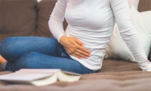
- Nonmenstrual pain in the lower abdomen. Their intensity depends on the stage of venous damage and the extent of the process. Grade 1 varicose veins of the pelvis are characterized by intermittent mild pain spreading to the lower back. In later stages it is felt in the abdomen, perineum and lower back and is prolonged and intense.
- Abundant mucous flow. The so-called leukorrhea does not have an unpleasant odor, does not change color, which would mean an infection. The volume of discharge increases in the second phase of the cycle.
- Increased symptoms of premenstrual syndrome and dysmenorrhea. Even before the onset of menstruation, the pain in women increases, until the onset of difficulty walking. During menstrual bleeding can become unbearable, spread to the entire pelvic area, perineum, lower back and even to the thighs.
- Another characteristic sign of varicose veins in the groin in women is discomfort during intercourse. It is felt in the vulva and vagina and is characterized by dull pain. It can be observed at the end of sexual intercourse. In addition, the disease is accompanied by increased anxiety, irritability and mood swings.
- As in the case of varicose veins of the pelvis in men, in the female part of the patients with such a diagnosis, the interest in sex gradually disappears. The cause of the dysfunction is both constant discomfort and decreased production of sex hormones. In some cases, infertility can occur.
Instrumental diagnostics
The diagnosis and treatment of varicose veins is performed by a phlebologist, vascular surgeon. Currently, the number of cases of VVP detection has increased due to new technologies. Patients with CPP are examined in several stages.
- The first stage is a routine examination by a gynecologist: taking a history, manual examination, ultrasound examination of the pelvic organs (to exclude other pathology). Based on the results, an additional examination by a proctologist, urologist, neurologist and other related specialists is prescribed.
- If the diagnosis is unclear but VVPT is suspected, pelvic vein ultrasound (USAS) is performed in the second stage. This is a non-invasive, highly informative screening method that is used in all women with suspected VVPT. If previously it was considered sufficient to examine only the pelvic organs (examination of the veins was considered difficult to access and optional), now at this stage ultrasound examination of the pelvic veins is a mandatory examination procedure. With the help of this method it is possible to determine the presence of varicose veins of the pelvis by measuring the diameters, the rate of blood flow in the veins and to determine in advance what is the leading pathogenetic mechanism - ovarian vein failure or venous obstruction. Also, this method is used for dynamic evaluation of conservative and surgical treatment of VVPT.
- The examination is performed transvaginally and transabdominally. Parametric veins, groin plexuses, and uterine veins are visualized transvaginally. According to various authors, the diameter of the vessels of these localizations varies from 2, 0 to 5, 0 mm (average 3, 9 ± 0, 5 mm), ie not more than 5 mm, and the average diameter of the arcuate veins is 1, 1 ± 0. 4 mm. Veins larger than 5 mm in diameter are considered dilated. The inferior vena cava, iliac veins, left renal vein, and ovarian veins are examined transabdominally to rule out thrombotic masses and extravasal compression. The length of the left renal vein is 6 to 10 mm, and its average width is 4 to 5 mm. Usually the left renal vein at the point where it passes over the aorta is somewhat flattened, but the reduction of its transverse diameter by 2-2, 5 times occurs without significant acceleration of blood flow, which ensures normal outflow without increasing pressure in the pretentious area. In the case of stenosis of a vein against the background of pathological compression, there is a significant reduction in its diameter - by 3, 5-4 times and acceleration of blood flow - over 100 cm / s. The sensitivity and specificity of this method are 78 and 100%, respectively.
- Examination of the ovarian veins is included in the mandatory examination of the pelvic veins. They are located along the anterior abdominal wall, along the rectus abdominis muscle, slightly lateral to the iliac veins and arteries. A sign of ovarian vein insufficiency in USAS is considered to be more than 5 mm in diameter with the presence of retrograde blood flow. For a complete examination, recurrence prevention and proper treatment tactics, ultrasound examination of the veins of the lower extremities, perineum, vulva, inner thigh and gluteal area should be performed.
- The development of medical technology has led to the use of new diagnostic methods. In the third stage, after ultrasound verification of the diagnosis, radiation diagnostic methods are used to confirm it.
- Pelvic phlebography with selective bilateral X-ray contrast ovaricography is one of the radiation invasive diagnostic methods that is performed only in a hospital setting. This method has long been considered the diagnostic "gold standard" for assessing dilatation and detecting valvular insufficiency in the pelvic veins. The essence of the method is the introduction of a contrast agent under the control of an X-ray installation through a catheter installed in one of the main veins (jugular, brachial or femoral) to the iliac, renal and ovarian veins. In this way it is possible to identify the anatomical variants of the structure of the ovarian veins, to determine the diameters of the gonadal and pelvic veins.
- The retrograde contrast of the gonadal veins at the height of the Valsalva test serves as a pathognomonic angiographic sign of their valvular insufficiency with visualization of abrupt dilation and corresponding curvature. This is the most accurate method for detecting May-Turner syndrome, postthrombophlebitic changes in the iliac and inferior vena cava.
- Compression of the left renal vein determines perirenal venous collaterals with retrograde blood flow in the gonadal veins, contrast stagnation in the renal vein. The method measures the pressure gradient between the left kidney and the inferior vena cava. Normal is 1 mm Hg. Art . ; gradient equal to 2 mm Hg. Art. , may offer light compression; with gradient>3 mm Hg. Art. can be diagnosed with aorto-mesenteric compression syndrome with hypertension in the left renal vein and gradient >5 mm Hg. Art. is considered hemodynamically significant stenosis of the left renal vein. Determining the pressure gradient is an important element of the diagnosis, because depending on its values, essentially different surgical interventions are planned on the veins of the pelvis, which is very important in modern conditions. Currently, this test (with a normal pressure gradient) can be used for therapeutic purposes - for embolization of ovarian veins.
- The next method of irradiation is emission computed tomography of the pelvic veins with in vitro labeled erythrocytes. It is characterized by the deposition of marked erythrocytes in the veins of the pelvis and visualization of the gonadal veins, allows to identify dilated splits of the pelvis and dilated ovarian veins in different positions, the degree of pelvic venous congestion, reflux of blood from the pelvic veins into the subcutaneousof the legs and perineum. Usually the ovarian veins are not contrasted, there is no accumulation of radiopharmaceuticals in the venous splits. To objectively assess the degree of venous congestion of the pelvis, the coefficient of pelvic venous congestion is calculated. But this method also has disadvantages: invasiveness, relatively low spatial resolution, inability to accurately determine the diameter of the veins, which is why it is currently not used so often in clinics.
- Video laparoscopic examination is a valuable tool for assessing the undiagnosed. In combination with other methods it can help determine the causes of pain and prescribe the right treatment. In varicose veins of the small pelvis in the area of the ovaries, along the round and wide ligaments of the uterus, the veins can be visualized in the form of cyanotic, dilated vessels with a thinned and tense wall. The use of this method is significantly limited by the following factors: the presence of retroperitoneal adipose tissue, the ability to assess varicose veins only in a limited area and the inability to determine reflux through the veins. At present, the use of this method is diagnostically justified in case of suspicion of multifocal pain. Laparoscopy allows you to visualize the causes of CPP, such as foci of endometriosis or adhesions, in 66% of cases.
Characteristics of the therapy
For the complete treatment of varicose veins of the pelvis, the woman must follow all the recommendations of the doctor, as well as change her lifestyle. First of all, you should pay attention to the loads, if they are excessively high, they should be reduced, if the patient leads an excessively sedentary lifestyle, it is necessary to exercise, to walk more often, etc.
Patients with varicose veins are strongly recommended to adjust their diet, to consume as little unhealthy food as possible (fried, smoked, sweet in large quantities, salty, etc. ), alcohol, caffeine. It is better to give preference to vegetables and fruits, dairy products, cereals.
Also, as a prevention of disease progression and for medical purposes, doctors prescribe the wearing of compression underwear for patients with varicose veins.
Medicines
ERCT therapy includes several important points:
- to get rid of the reverse flow of venous blood;
- relieving the symptoms of the disease;
- stabilization of vascular tone;
- improving blood circulation in the tissues.
Varicose veins preparations should be taken in courses. Other drugs that act as painkillers can be taken only during a painful attack. For effective therapy, the doctor often prescribes the following drugs:
- phleboprotectors;
- enzyme preparations;
- drugs that relieve inflammatory processes with varicose veins;
- pills to improve blood circulation.
Surgery
It is worth acknowledging that conservative methods of treatment give really visible results mainly in the initial stages of varicose veins. At the same time, the problem can be fundamentally solved and the disease can be completely eliminated only through surgery. In modern medicine there are many options for surgical treatment of varicose veins, consider the most common and effective types of operations:
- ovarian embolization in the ovaries;
- sclerotherapy;
- uterine ligament plasticity;
- removal of varicose veins by laparoscopy;
- compression of veins in the small pelvis with special medical clips (clipping);
- crossectomy - ligation of veins (prescribed if, in addition to the pelvic organs, the vessels of the lower extremities are also affected).
During pregnancy, only symptomatic treatment of varicose veins of the pelvis is possible. We recommend wearing compression tights, taking phlebotonics on the recommendation of a vascular surgeon. During the II-III trimester, phlebosclerosis of the varicose veins of the perineum can occur. If due to varicose veins there is a high risk of bleeding in spontaneous birth, the choice is made in favor of operative birth.
Physiotherapy
The system of physical activity for the treatment of varicose veins in women consists of exercises:
- "Bicycle". We lie on our backs, throw our hands behind our heads or place them along our body. Raising our legs, we make circular movements with them, as if we were pedaling a bicycle.
- "Birch". We sit face up on any hard, comfortable surface. Lift your legs up and start them slightly behind your head. Supporting the lumbar region with your hands and placing your elbows on the floor, slowly straighten your legs, lifting your body up.
- "Scissors". The starting position is on the back. Raise your closed legs slightly above floor level. We spread the lower limbs to the side, put them back and repeat.
Possible complications
Why are varicose veins of the pelvis dangerous? The following effects of the disease are often recorded:
- inflammation of the uterus, its appendages;
- uterine bleeding;
- abnormalities in the work of the bladder;
- the formation of venous thrombosis (small percentage).
Prevention
In order for the varicose veins in the small pelvis to disappear as soon as possible and in the future there will be no recurrence of the pathology of the pelvic organs, it is worth adhering to simple preventive rules:
- perform gymnastic exercises daily;
- prevention of constipation;
- follow a diet in which plant fiber should be present;
- do not stay in one position for a long time;
- take a contrast shower on the perineum;
- to prevent varicose veins, it is better to wear extremely comfortable shoes and clothes.
Preventive measures aimed at reducing the risk of the appearance and progression of varicose veins in the pelvis are mainly reduced to the normalization of lifestyle.